MODULE 8 - VIEWS-ALL
The
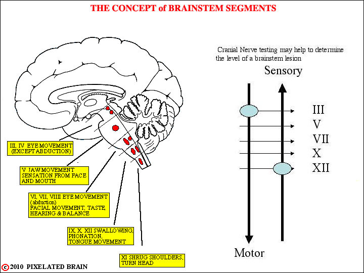
FIGURE 8-1
The sensory and motor pathways, ascending or descending through the brainstem, are obliged to thread their way through a region littered with cranial nerve motor and sensory nuclei. Damage to a small region where a pathway is close to a nucleus or exiting cranial nerve creates a unique combination of defects, or a syndrome. Knowing the anatomy thus often allows one to localize lesions with great precision.
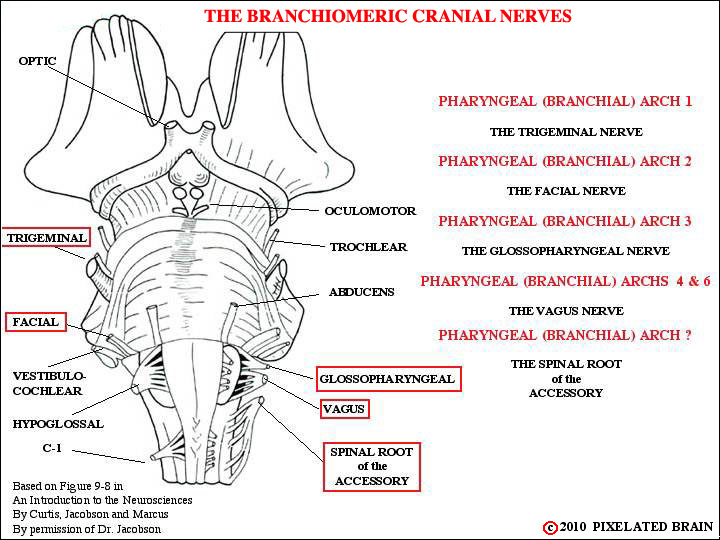
FIGURE 8-2
If the concept of branchial arches is new to you, see arch 1 for a summary. The fifth arch exists only briefly during development, and since no permanent structures are derived from it, we ignore its brief existence. There is a debate about the classification of the spinal root of the accessory nerve as a branchiomeric one, because there is no seventh arch. We discuss the arguments pro and con when we consider the nerve in more detail in Figure 8-13.
When we first treated cranial nerves in Module 2, we told you - in Fig. 2-14 - not to worry about the details. We said we'd do that in Modules 8 and 9. Well, Module 8 is here and the time for details has come. For starters, look at Figure 1-5 and - for the 5 cranial nerves listed above - make certain you can draw a circle around the opening through which the nerve passes to exit from the skull. If you need help, consult Blumenfeld's Table 12.2.
This view gives us a chance to drive home one of the points made in the preceding view. In your mind, try to draw in the descending pyramidal tract on this view. Note that it passes close to three cranial nerves - the oculomotor, the abducens and the hypoglossal. These relationships give rise to three clinical syndromes - the so - called "alternating hemiplegias". In each case the motor loss is on the contralateral side of the body, and it is associated with an ipsilateral impairment in cranial nerve function.
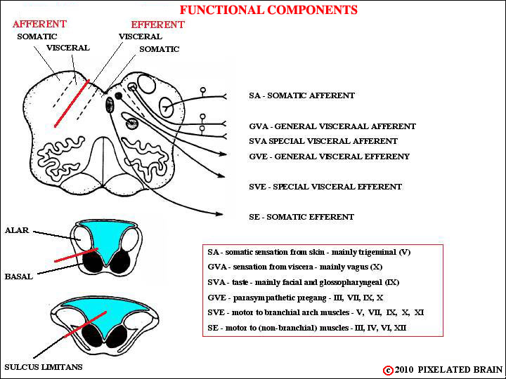
FIGURE 8-3
You will remember (we hope) that when the neural plate rolled up to form a tube the sulcas limitans served to form a marker, seperating the dorsal "alar plate" from the ventral "basal plate" as seen in emb_fig 06 . There are, however, regions where the lips of the neural plate don't meet; in these regions the roof of the central cavity is sealed by choroid plexus tissue, and the alar plate tends to lie lateral to the basal plate, rather than dorsal to it - as shown above and in emb_fig 08 . The picture is further complicated by the appearance of four new functional groupings, serving functions unique to the head. These are thought of as "visceral" because the are associated with the rostral end of the gut or alimentary canal. As the diagrams show, the sensory nuclei lie dorsolateral to the sulcus limitans and the motor nuclei lie ventromedial to it.
The sulcus limitans is only obvious in the caudal medulla, but a suggestion of it is also present in the diencephalon where a slight "valley" separates the dorsal thalamus (which might be thought of as a sensory structure) from the ventrally placed hypothalamus (which might be thought of as a motor structure).
Blumenfeld illustrates this topic it in his Figure 12.4.
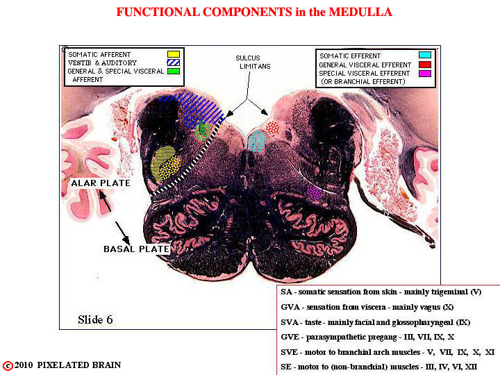
FIGURE 8-4
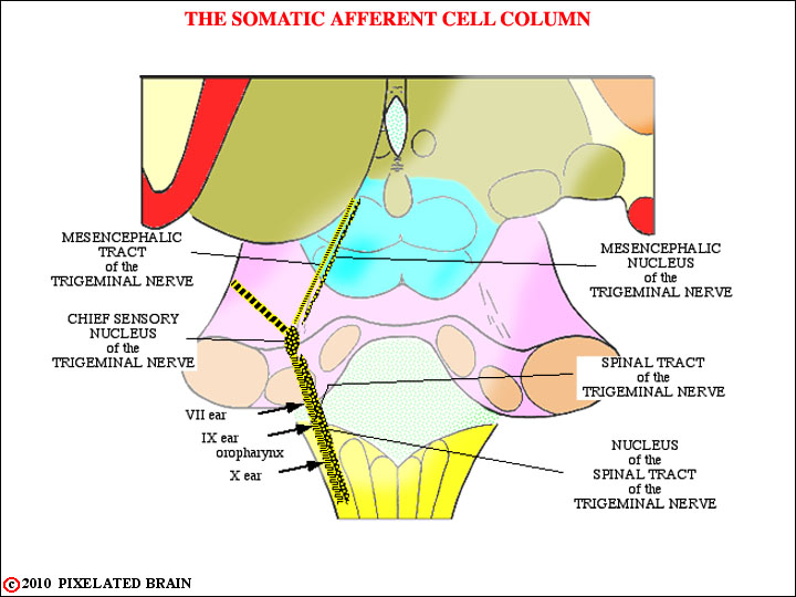
FIGURE 8-5
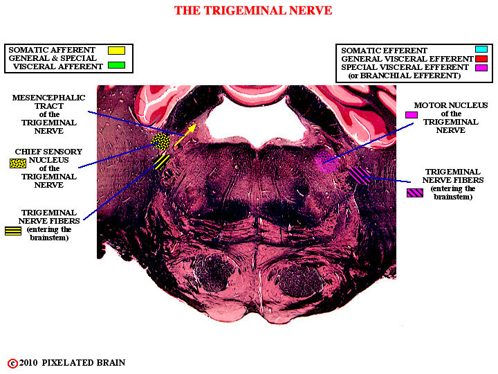
(General) somatic sensory function for the face is served primarily by the trigeminal nerve and we considered these structures in detail in Module 4. The view above is just a schematic version of Figure 4-4.
Something we didn't tell you earlier is that during development all the branchial arches share a common turf in the region aroind the ear. Thus cranial nerves 7, 9 and 10 each have a few somatic sensory fibers. For more detail, see descriptions of the individual nerves, later in the module.
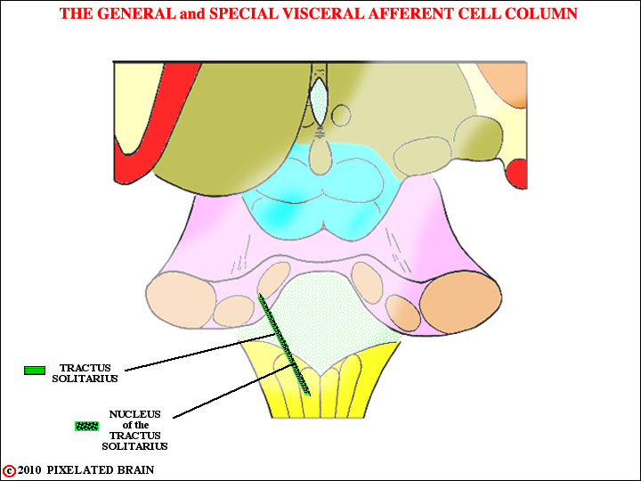
FIGURE 8-6
Axons of cranial nerves VII, IX and X conveying the sense of taste (SVA) enter the brainstem within their respective nerves and travel upward in the Tractus Solitarius to terminate in the most rostral part of the Nucleus of the Tractus Solitarius, sometimes known as the gustatory nucleus. Second order neurons within the nucleus send fibers rostrally to terminate in the VPM nucleus of the thalamus. The pathway is probably bilateral, and runs near the ventral secondary trigeminal tract (Blumenfeld places it in the central tegmental tract). From the thalamus, the pathway ascends to a cortical region terminating laterally, near the tongue area of the postcentral gyrus and adjacent to the insula.
Axons of cranial nerves IX and X conveying general sensation (GVA) from the viscera enter the brainstem and travel downward in the Tractus Solitarius to terminate in the caudal part of the nucleus. We are not consciously aware of these sensations. Neurons in this part of the nucleus send axons to terminate in a broad array of brainstem nuclei (the Dorsal Motor Nucleus of the Vagus, the Nucleu Ambiguus, the Reticular Formation) , thus participating in a variety of visceral and autonomic reflexes. Fibers also pass rostrally to provide an input to the Hypothalamus and Amygdala.
If you want to have a little fun, try the QUIZ.
Most books do not include the parabrachial nucleus in the taste pathway. It lies adjacent to the superior cerebellar peduncle (Brachium conjuntivum - hence, the name) in the rostral pons.
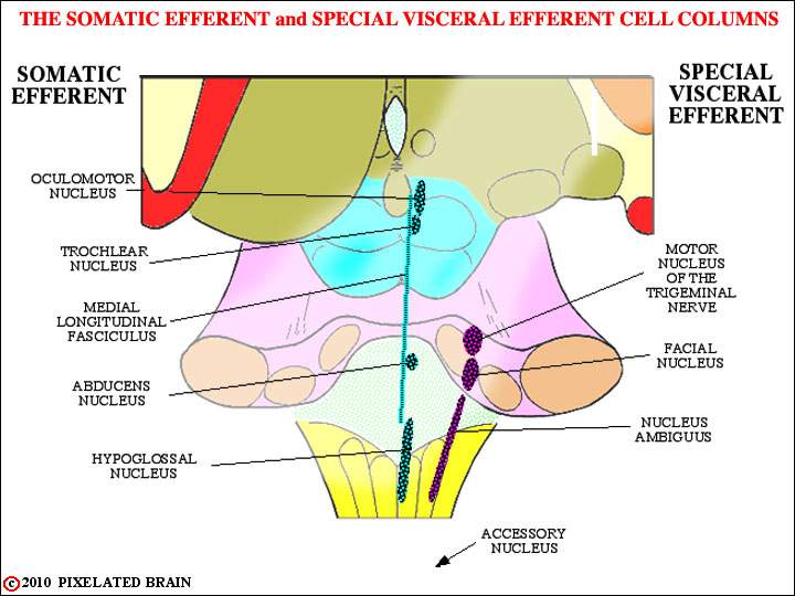
FIGURE 8-7
The oculomotor, trochlear and abducens nerves innervate the muscles that move the eyes. The medial longitudinal fasciculus serves to connect these nuclei and coordinate their activity. The hypoglossal nerve, of course, has nothing to do with eye movement - rather, it innervates the muscles of the tongue. These nuclei are all a part of the Somatic Efferent (SE) cell column and we take them up in Module 9
The four nucle of the Special Visceral Efferent (SVE) cell column are shown here in purple. The (spinal) Accessory nucleus lies in the upper 5 or 6 segments of the cord, indicated here by the arrow. While most of the cell columns, like the SE one above are at best, interrupted ones, the SVE column is really an almost continuous one. The motor neurons in these nuclei are typical "lower motor neurons", just like those in the spinal cord. What makes them "special" is that they innervate muscles of branchial arch origin. The muscles are striated, so many people are uneasy about calling them "special VISCERAL motor. To get around this they are often called Branchiomotor.
Just like spinal motor neurons, the SVE ones are driven by upper motor neurons in the lateral part of the precentral gyrus. This pathway departs from the corticospinal one at the midbrain level (Figure 5-5), so we can't call it part of the pyramidal tract (it doesn't go through the pyramids of the medulla). It is called the coriticobulbar pathway and you traced it in Module 5 (remember all those yellow dots). If you want a quick review, click on Slide 28, and then use the "caudal buttons" to follow it downward through the tegmentum. We discuss it later in the module.
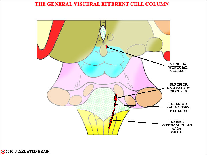
FIGURE 8-8
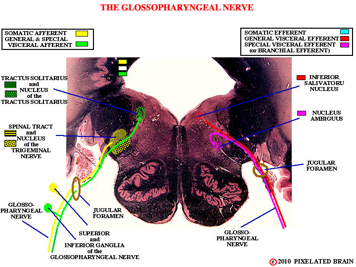
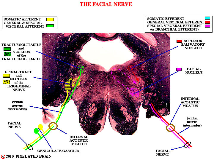
As you know, the autonomic nervous system is made up of two parts - the sympathetic (thoracolumbar) system and the parasympathetic (craniosacral). Both systems have 2 neuron pathways , involving preganglionic and postganglionic neurons. Here , we are looking at the cranial part of the parasympathetic nervous system, and the nuclei you see contain preganglionic neurons. The neurons in the Superior Salivatory Nucleus will send axons out in the Facial Nerve: the ones in the Inferior Salivatory Nucleus contribute axons to the Glossopharyngeal Nerve, and the ones in the Dorsal Motor Nucleus of the Vagus contribute axons to the Vagus Nerve.
We shouldn't forget the Nucleus of Edinger-Westphal. It contributes parasympathetic axons to the oculomotor nerve... yes, it's not a branchiomeric nerve, but it does have parasympathetic fibers in it. Go figure!
For more information about these GVE (parasympathetic) fibers, see the views of the individual cranial nerves.
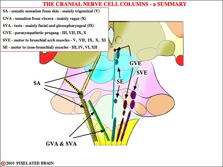
FIGURE 8-9
Use this view to test your short term memory. Point to each of the nuclei, one by one and ignoring the SE column, see if you can name it and have some idea about what it does, then click the next button to check your answers
To call up slides click on FIGURE 8-24 .
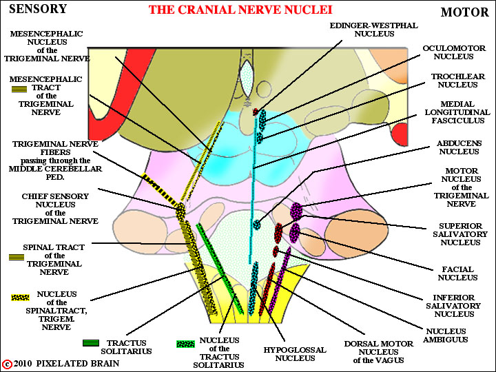
FIGURE 8-10
Well, we didn't want to show you this view at the start, because we figured you'd never get past the first slide. Most books have views of this sort .. see, for example, Blumenfeld's Figure 12.5 or Kandel's Figure 44.6. The thing that makes our view special is that it was constructed to scale from a set of slides cut through a real brainstem ....the slides we call the Cu Series. You can check it out by calling up FIGURE 8-24 and looking at specific slides in the series.
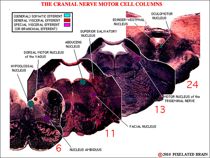
FIGURE 8-11
The idea here was to try to demonstrate that the cell columns really are columns, but it isn't easy to do on a limited number of sections. Here we are looking at the motor columns and you may be able to see that:
1) The Nucleus Ambiguus, Facial Nucleus and Trigeminal Motor Nucleus (SVE) do line up, one in front of the other (but difficult to pick out because of the colors used)
2) The Dorsal Motor Nucleus of the Vagus, the Superior Salivatory Nucleus and the Edinger-Westphal Nucleus (GVE) sort of line up, but it is a very discontinuous cell column. (The Inferior Salivatory Nucleus isn't present on the slides selected).
3) The Hypoglossal Nucleus, the Abducens Nucleus and the Oculomotor Nucleus (SE) line up, but again the column is a discontinuous one. (The Trochlear Nucleus isn't present on the slides selected).
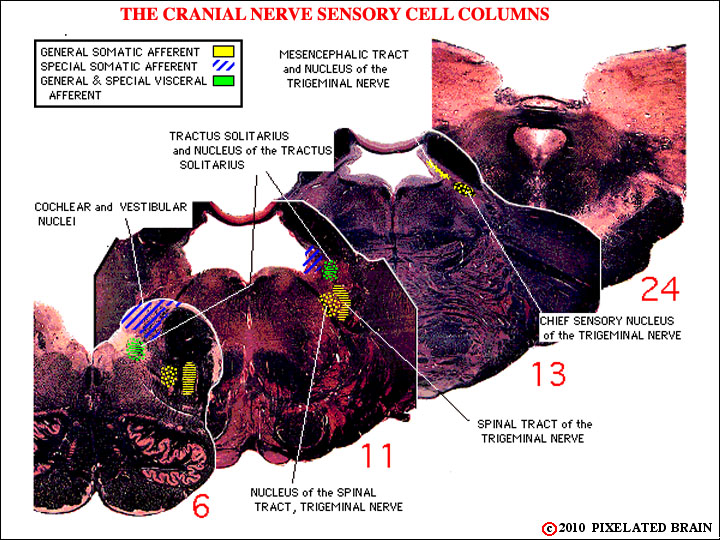
FIGURE 8-12
This view is of the sensory columns. We have slipped up, as we said we might, and called the Vestibular and Auditory Nuclei "Special Somatic Afferent". Try to ignore this.
The idea here is just to show that the Nucleus of the Tractus Solitarius is a column (admittedly, a short one) that is present in slides 6 and 11, and that the trigeminal nuclei form a column that extends from the spinal cord (not shown) as far rostrally as slide 13 (and beyond).
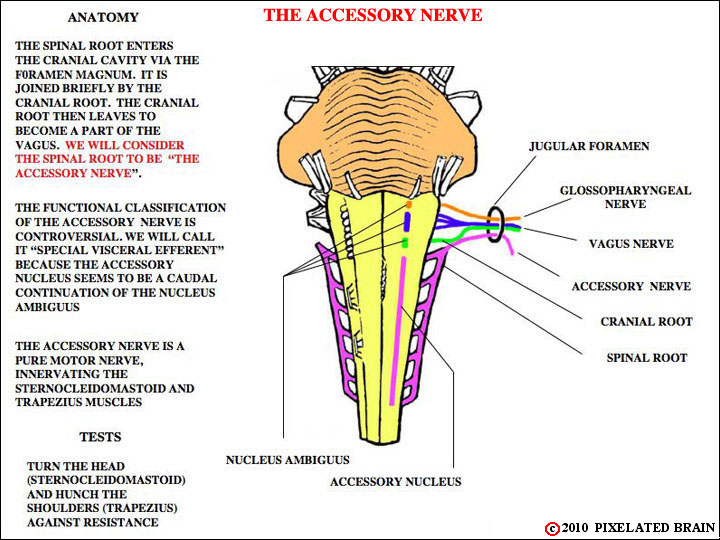
FIGURE 8-13
You have seen the accessory nerve before, in views 2-16, 2-41, 2-42 and 2-43. Another nice view of the real thing is gluhb_01_3.html , but it is not labeled in that view.
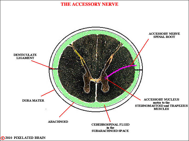
FIGURE 8-14
OK, so here's a trick question for you to use on your friends at Medical School X. "What spinal cord lower motor neurons send their axons out of the cord without passing through a ventral root?"
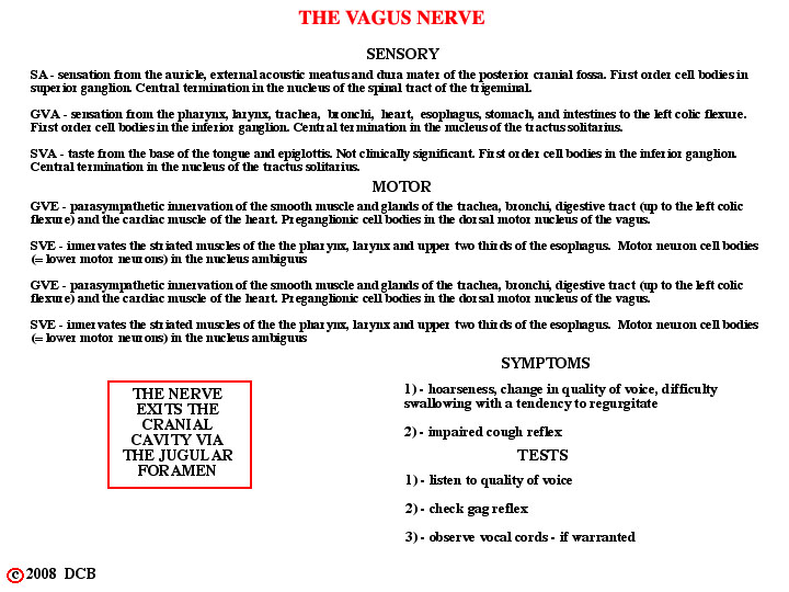
FIGURE 8-15
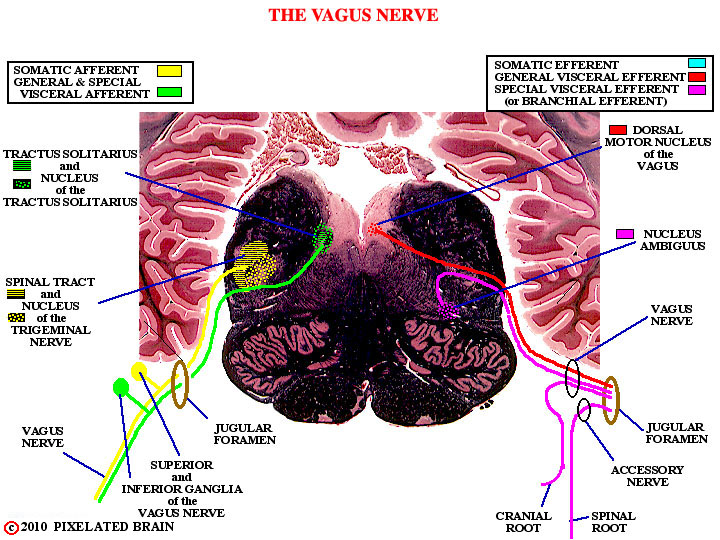
We should have included chemo and baroreceptors from the aortic arch in the GVA list. All these GVA fibers terminate in the caudal part of the Nucleus of the Tractus Solitarius.
For views of the exiting Vagus Nerve, see Figure 2-16, Figure 2-39, Figure 2-41 and Figure 2-42.
FIGURE
z
FIGURE 8-17
FIGURE
z
FIGURE 8-19
FIGURE
z
FIGURE 8-21
FIGURE 8-22
FIGURE
z
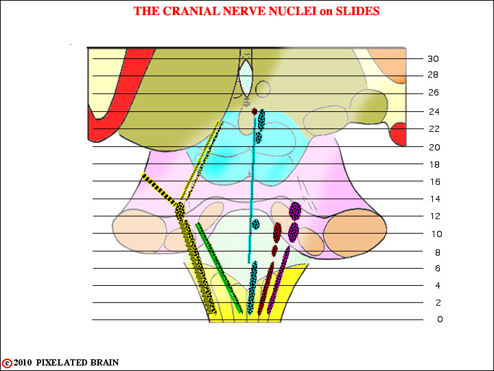
FIGURE 8-24
To look at similar view in which the cell columns are named click on Figure 8.9.
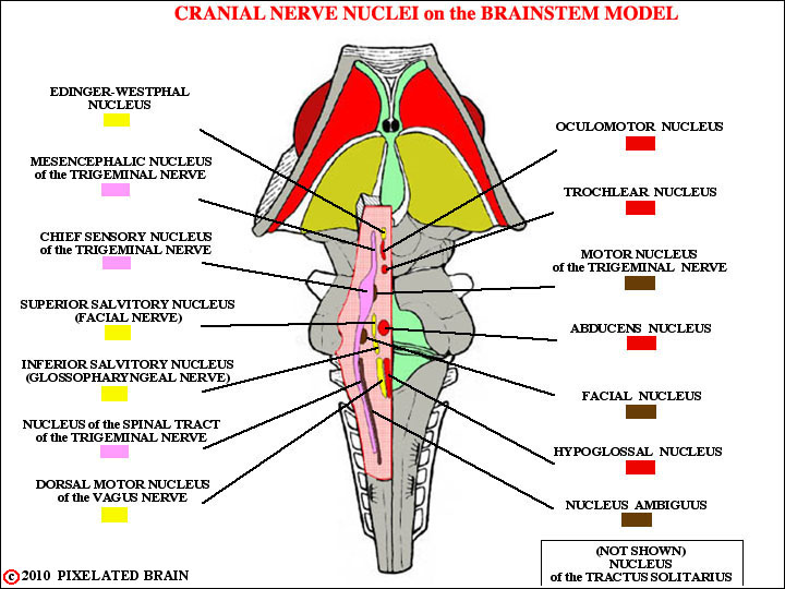
FIGURE 8-25
Compare with Fig. 1-13, Fig. 1-15, Fig. 1-16, Fig. -17, Fig. 1-18,
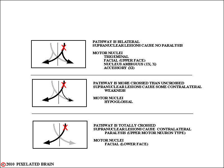
FIGURE 8-26
The point about the effect of supranuclear lesions (ones damaging the corticobulbar pathway) on the function of the facial and hypoglossal nerves is an extremely important one. To be certain you are on it read pages 482 - 4 in Blumenfeld and check out Figure 12.13.
The terse comment at the bottom of the box, "but the sternocleidomastoid is special" refers to the unusual action of this muscle. Generally we recognize that each hemisphere is concerned with the opposite side of the body and - by extension - the opposite half of surrounding space. Thus, stimulation of the appropriate part of the right cerebral hemisphere will cause both the head and the eyes to turn to the left. But it is the right sternocleidomastoid muscle that helps turn the head to the left by pulling the mastoid process forward. So, most authors (Kandel, Blumenfeld (Pg 499) and Wilson-Pauwels et al - the yellow cranial nerve book folks) say that the corticobulbar input to the part of the accessory nucleus innervating the sternocleidomastoid muscle is uncrossed....ie the right hemisphere controls the right sternocleidomastoid muscle.
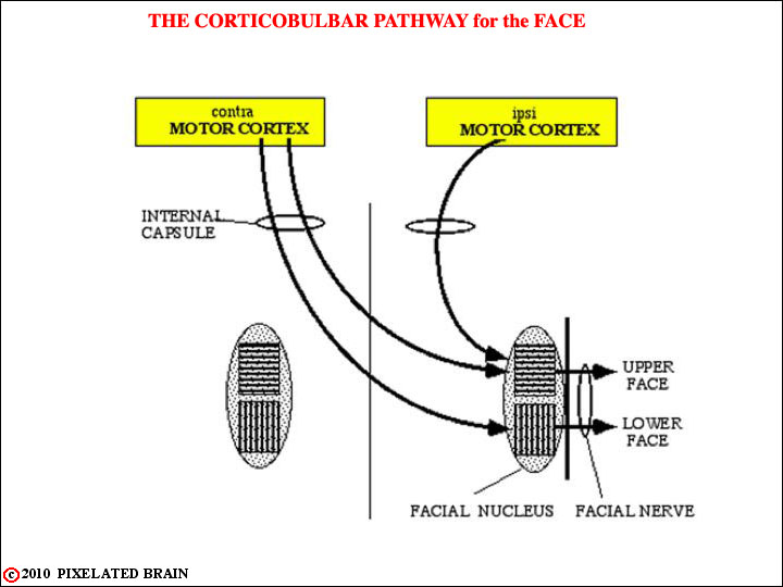
FIGURE 8-27
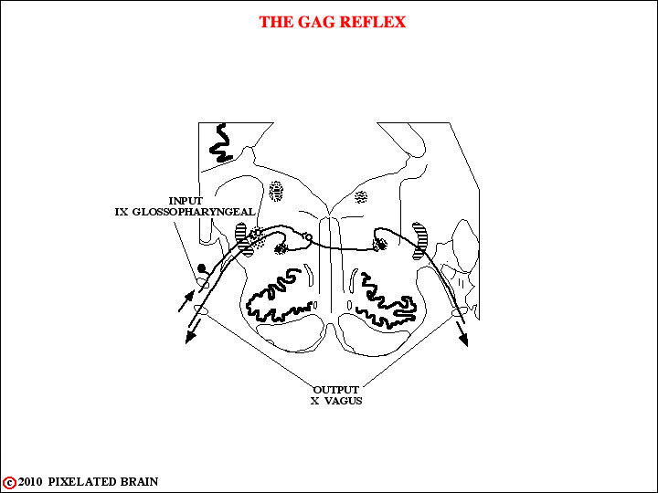
FIGURE 8-28
FIGURE 8-29
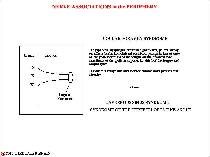
FIGURE 8-30
FIGURE
z
FIGURE
z