MODULE 8 - SECTION 4 - THE BRAINSTEM SLIDE SERIES
Your final job is to look through the brainstem slide series (C-1, C-2, Transition, and 1-27) and identify the various nuclei and tracts that relate to the cranial nerves. Go through the slides in order, or skip around - whichever works best for you. The slides are presented in order below, or you can use Figure 8-24 to navigate through them.
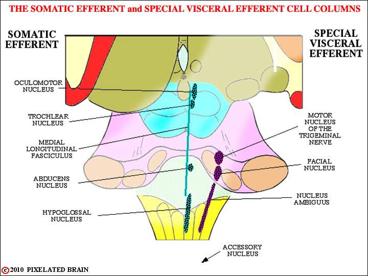
Note that in this slide series we have included the nuclei of the somatic efferent cell column, even though we don't take them up in this module.
- - - If you are having trouble visualizing where all these nuclei are in the real brain, you might try looking at the brainstem model. The dorsal surface of the brainstem has been "dissected away" to reveal most of the nuclei. Open Figure 8-25 in another window and use it as a guide. See how many of the nuclei you can find.
After the slides, be sure to continue reading below about THE CORTICOBULBAR PATHWAYS, BRAINSTEM REFLEXES, and NERVE ASSOCIATIONS IN THE PERIPHERY.
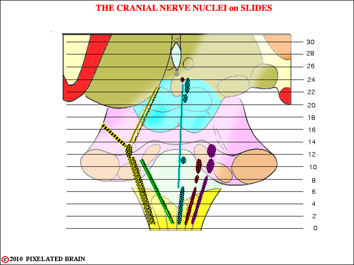
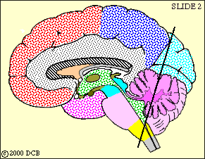
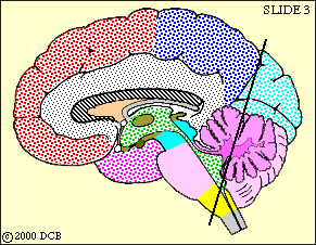
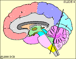
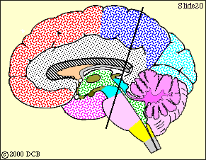
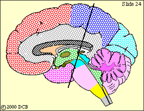
The planes for our slides.


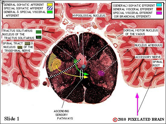
Cranial Nerves on Slide 1
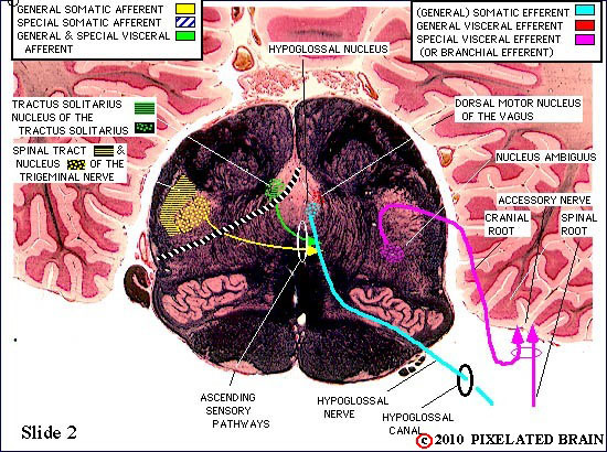
Cranial Nerves on Slide 2
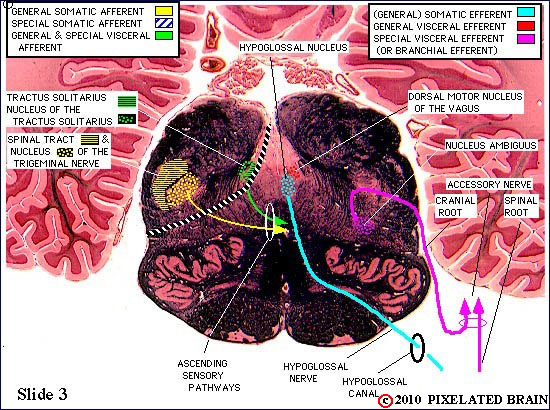
Cranial Nerves on Slide 3
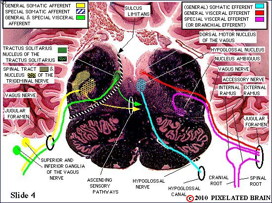
Cranial Nerves on Slide 4
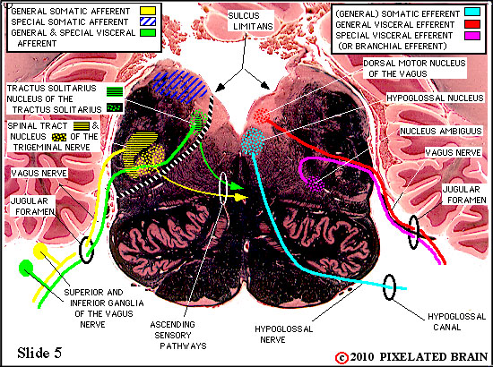

Cranial Nerves on Slide 5
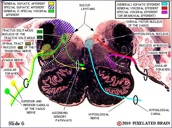
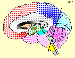
Cranial Nerves on Slide 6

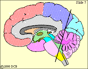
Cranial Nerves on Slide 7


Cranial Nerves on Slide 8


Cranial Nerves on Slide 9


Cranial Nerves on Slide 10
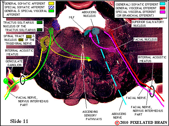
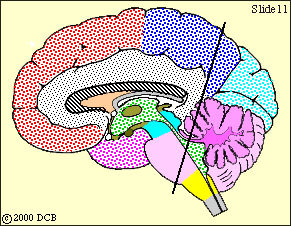
Cranial Nerves on Slide 11


Cranial Nerves on Slide 12


Cranial Nerves on Slide 13


Cranial Nerves on Slide 14


Cranial Nerves on Slide 15
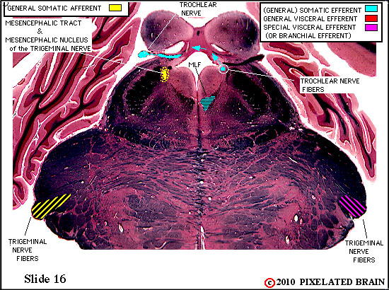

Cranial Nerves on Slide 16


Cranial Nerves on Slide 17
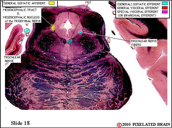

Cranial Nerves on Slide 18
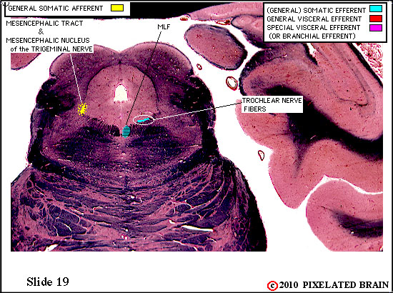

Cranial Nerves on Slide 19
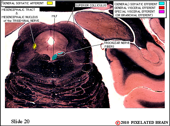
Cranial Nerves on Slide 20
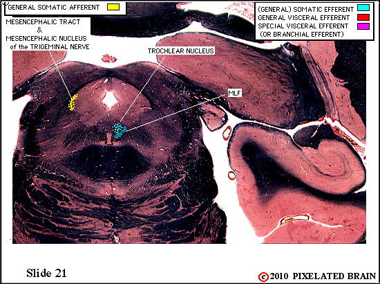

Cranial Nerves on Slide 21
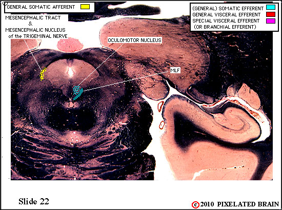

Cranial Nerves on Slide 22


Cranial Nerves on Slide 23
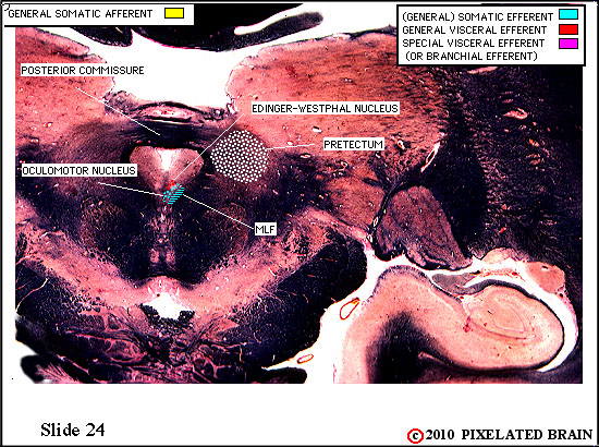
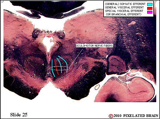
Cranial Nerves on Slide 24

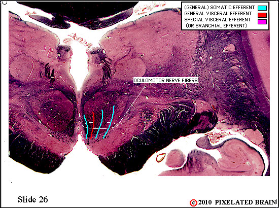
Cranial Nerves on Slide 25

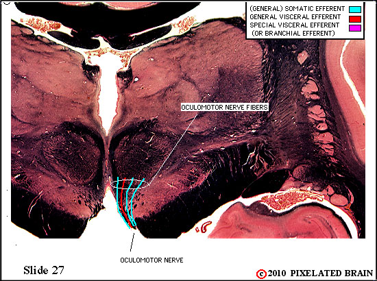
Cranial Nerves on Slide 26
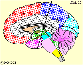
Cranial Nerves on Slide 27
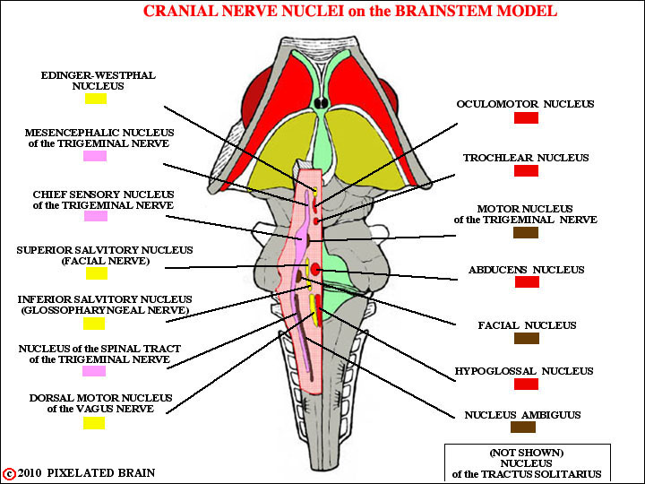
If you are having trouble visualizing where all these nuclei are in the real brain, you might try looking at the brainstem model. The dorsal surface of the brainstem has been "dissected away" to reveal most of the nuclei. Using this view as a guide, see how many of the nuclei you can find.
32
THREE MORE POINTS
THE CORTICOBULBAR PATHWAYS
Most motor nuclei of the brainstem are collections of typical "lower motor neurons". They receive a descending input from "upper motor neurons" in the lateral part of the precentral gyrus. Nuclei in this group include all the special visceral efferent nuclei and the hypoglossal nucleus .
THE CORTICOBULBAR PATHWAYS
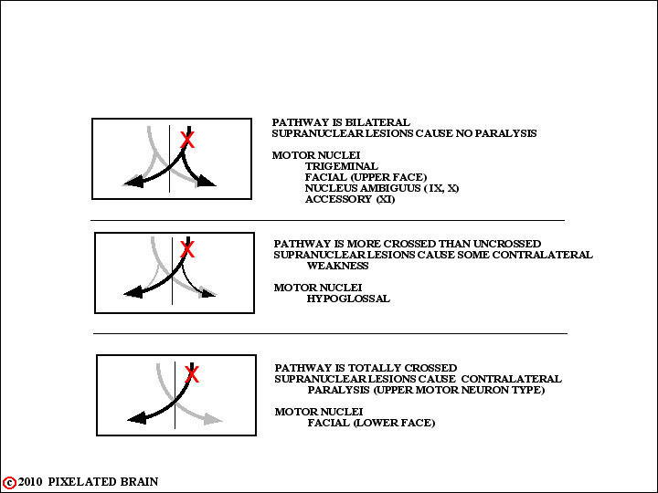
Unlike the situation in the spinal cord the descending input to these nuclei tends to be bilateral, but there are important exceptions. This view summarizes the situation.
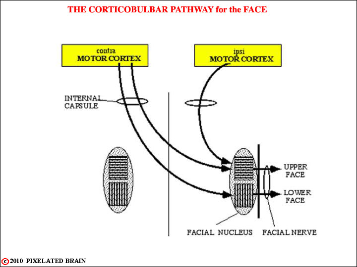
THE CORTICOBULBAR PATHWAYS
The case of the corticobulbar input to the facial nucleus is of particular note.
This view shows the cortical input in schematic fashion.
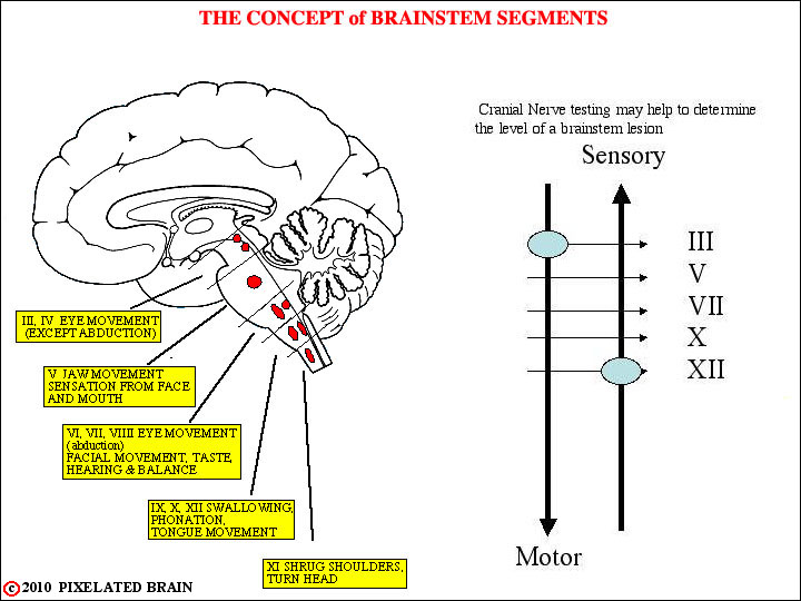
BRAINSTEM REFLEXES
As you know, a variety of reflexes are used to test the integrity of spinal cord segments. The same principle applies to the brainstem, and this view attempts to make this point.
BRAINSTEM REFLEXES
To give an obvious example, the trigeminal nerve - in a sense - "owns" the mid pons. Thus, if the trigeminal is functioning normally, it suggests (but doesn't prove) that there are no major problems in this segment of the brainstem. One way to check on the nerve is to test the jaw jerk. This view will remind you of the the anatomical basis for this monosynaptic reflex.
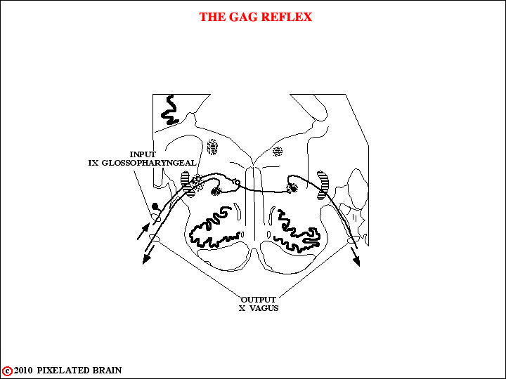
BRAINSTEM REFLEXES
There are several other reflexes of this sort that you should be familiar with. One, the gag reflex, is shown here.
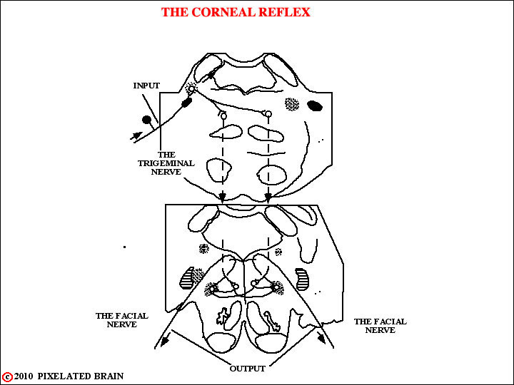
BRAINSTEM REFLEXES
The anatomy of a second, the corneal reflex, is a bit more complicated since the afferent limb utilizes the trigeminal nerve and the efferent limb utilizes the facial nerve - as shown here. If the corneal reflex is impaired, then the first task is to determine which of these cranial nerves is at fault. As Blumenfeld points out on Page 484, this reflex is also influenced by other regions of the brain.
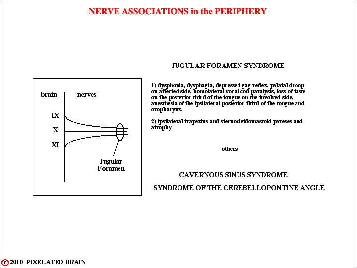
NERVE ASSOCIATIONS IN THE PERIPHERY
We already know that many brainstem syndrome are based on the fact that structures with unrelated functions just happen to be placed next to each other.
Thus a small lesion may damage all of them and give a characteristic clinical picture.
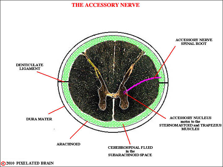
The same principle applies to the peripheral nervous system. One example is the Jugular Foramen Syndrome. Since three cranial nerves, the glossopharyngeal, the vagus and the accessory, squeeze their way out of the cranial cavity through the jugular foramen, a small lesion here gives the result described in this figure.
See if, based on your knowledge of the anatomy, you can come up with a good description of the Cavernous Sinus Syndrome and the Syndrome of the Cerebellopontine Angle.