MODULE 1 - SECTION 6 - INTERNAL ANATOMY of the CEREBRAL HEMISPHERE
One of the most difficult topics for students of neuroanatomy to master has to do with the internal anatomy of the cerebral hemispheres. There are actually only a few structures (and names) involved, but the manner in which the brain has developed has caused the relationships between them to be complex. Added to this is the fact that the most convenient way to study the hemisphere involves the use of serial sections - and that no matter what plane of section one picks, the cuts will pass through some structures at odd angles. The end result for the student is confusion which usually does not begin to lift until the last weeks of the course. We hope that by focusing on this subject at the start we will help you avoid this. But, be prepared for some tough going!
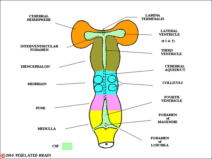
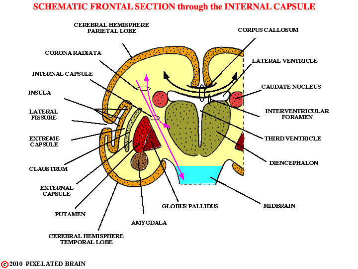
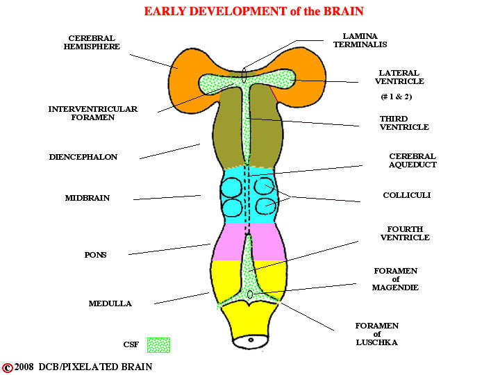
Early in the module we felt obliged to say a little about brain development because we wanted to explain how the ventricular system came to be. So, when we last looked at the developing hemisphere, in this figure, it was just a blister, extending laterally from the diencephalon. In fact, things are more complicated, as shown next, in a schematic frontal section through the hemisphere and diencephalon.
A sheet of cells, the cerebral cortex, lies just below the cortical surface. Underneath the cortex lies the "white matter" - actually a jumble of axon bundles running every which-way.
Buried within the white matter are several nuclei - each of them a region of densely packed neuron cell bodies. As the axons of the white matter try to pass to and from the cortex, and from one cortical region or hemisphere to another, they must pass between and around these so-called deep nuclei. The nuclei are, in a sense, the troublemakers; as a group, they are known as the basal ganglia and they include the caudate nucleus, the putamen, the globus pallidus, the amygdala and the claustrum.
The major white matter pathways threading between the nuclei include:
1) the corpus callosum, connecting one cerebral hemisphere with the other and often forming the roof of the lateral ventricle.
2) the internal capsule, which contains some fibers (axons) interconnecting the diencephalon with the cerebral cortex and others which pass from the cortex to more caudal parts of the brainstem and the spinal cord.
3) the corona radiata, which represents the extension of the internal capsule toward the surface of the hemisphere, into the region where the fibers are no longer trapped between the caudate and the lentiform nucleus.
4) the external and extreme capsules, pathways connecting one part of the hemisphere with another and passing on either side of the claustrum.
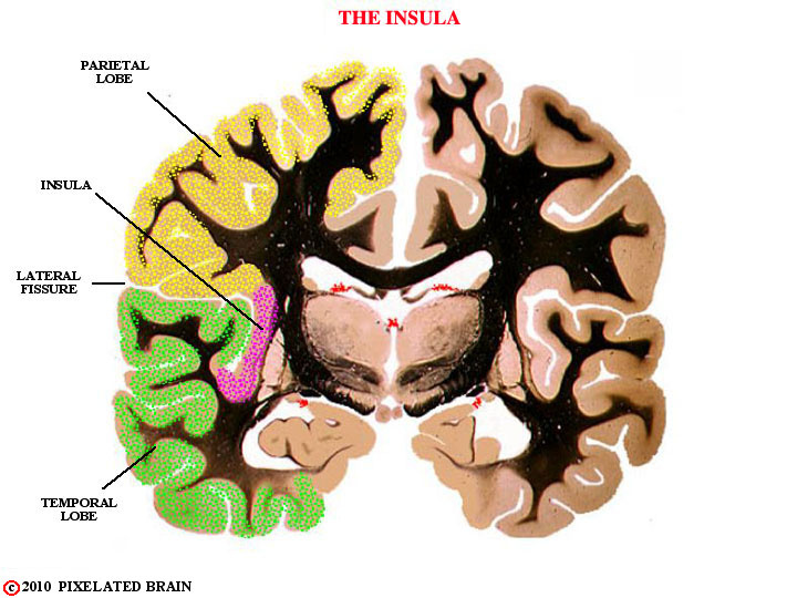
Note that the myelinated axons are colored yellow in the previous figure, but they give a white appearance to the region in fresh brain specimens. Because mylein stains black, the white matter appears black in histological sections through the brain, such as the one shown here.
THE BRAIN DEVELOPS...
If the brain remained as depicted, then the internal anatomy of the hemisphere would not be difficult to visualize, BUT

THE BRAIN DEVELOPS...
the fact is that as the hemisphere enlarges, it extends back over the diencephalon and midbrain, with part of it then pushing forward again to form the temporal lobe.

The results of this development include the next views.
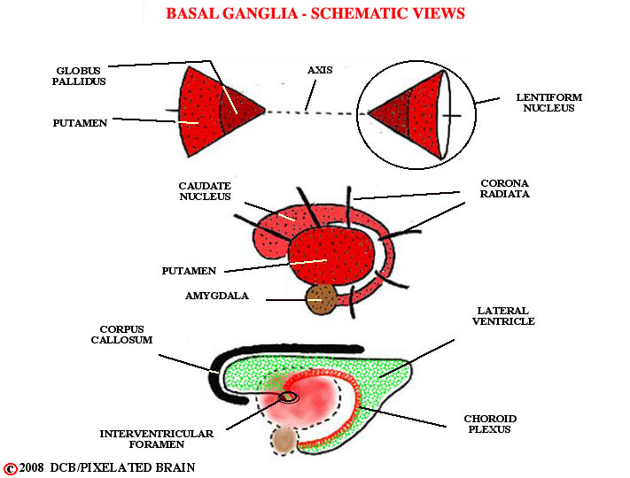
The lentiform nucleus resembles a cone lying on its side. The apex of the cone faces medially and is formed by the globus pallidus. The base of the cone faces laterally and is formed by the putamen. The cone, itself, does not take part in the migration associated with the development of the hemisphere. Rather, it remains fixed, centrally, and other structures circle it.
For a gross view see DiganatA_2.2. The surface of the posterior part of the putamen is covered by the external capsule.
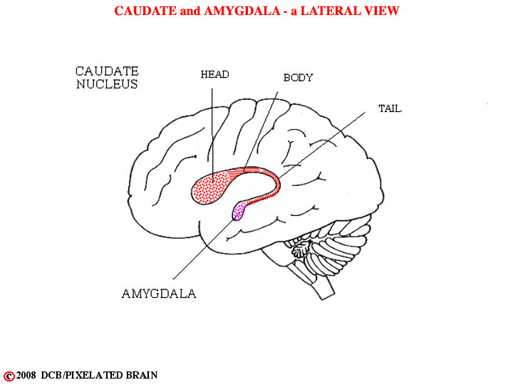
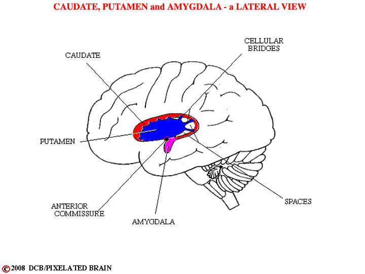
The caudate nucleus has a long tail (hence, its name) which is drawn out into the temporal lobe. It ends there by "bumping into" another basal ganglia nucleus - the amygdala. The amygdala is also in direct contact dorsally with the ventral edge of the putamen, as shown in the next slide.
The axis of the lentiform nucleus passes through the region where the lateral ventricle communicates with the third ventricle, i.e., the axis goes through the inter ventricular foramen of Monro. The lentiform nucleus remains centrally positioned, and the caudate "wraps around it" as it migrates into the temporal lobe.
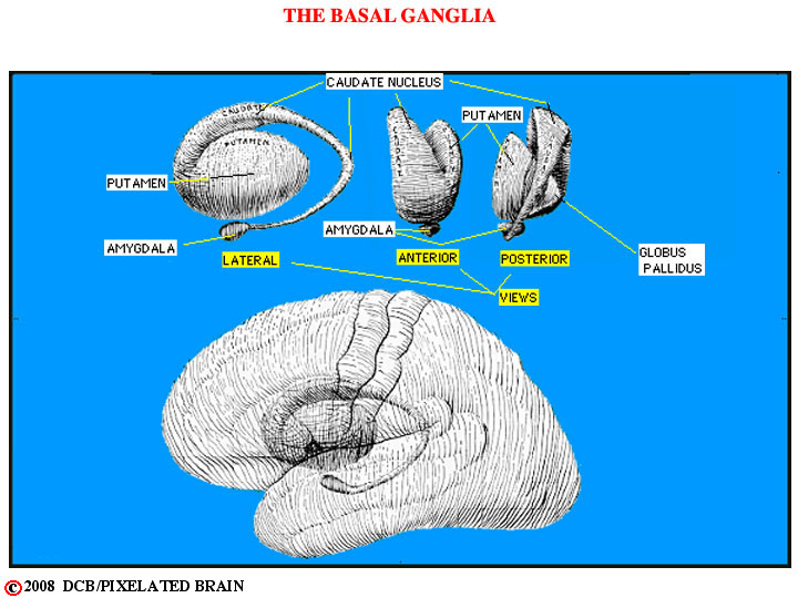
Shows the interrelationships between basal ganglia nuclei
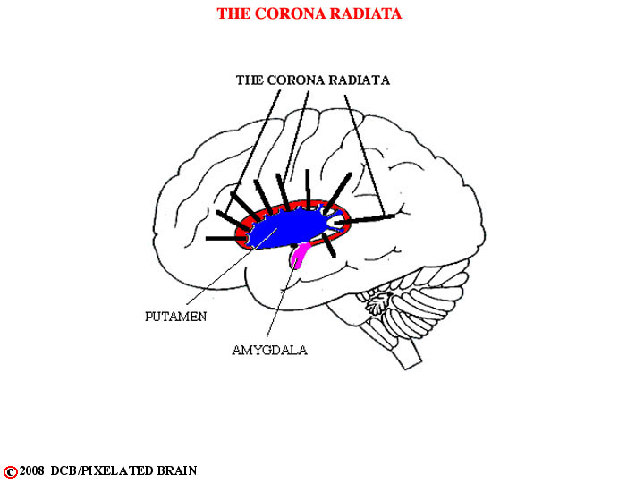
Since the fibers (axons) of the internal capsule pass between the caudate and the lentiform nucleus, they are drawn out into a fan-shaped array as the caudate heads for the temporal lobe. The fact that these fibers, in lateral view, resemble the rays of the sun radiating from behind the moon (lentiform nucleus) at the time of an eclipse accounts for the name corona radiata.
For a gross view see DiganatA_2.4 (putamen still there) and DiganatA_2.5 (putamen and globus pallidus removed.
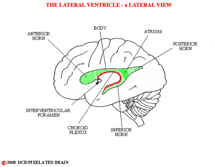
The lateral ventricle retains its relationship to the caudate nucleus during development (note that the caudate always forms part of the wall of the ventricle) with the result that it is also drawn out into the temporal lobe, bringing choroid plexus along. Additional extensions of the lateral ventricle into frontal and occipital lobes are blind evaginations with no defect in the wall - hence, no choroid plexus.
Note that because the lentiform nucleus resembles a cone, it will appear triangular in both coronal (frontal) and horizontal sections through the brain ; it will be circular in parasagittal sections. Because of the curved shape of both the caudate nucleus and the lateral ventricle, they may be cut twice in coronal sections and, more rarely, in horizontal sections.
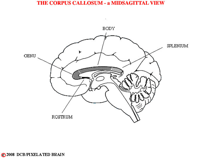
The process of migration also elongates the corpus callosum.
- In summary, deep within the cerebral hemisphere lie a group of nuclei (nuclei = groups of neuronal cell bodies packed together) called the basal ganglia. These nuclei include the caudate, the putamen, the globus pallidus, the amygdala and the claustrum. The migration of neural tissue, as the hemisphere has enlarged, has caused these nuclei (particularly the caudate and the associated lateral ventricle) to assume odd shapes. Threading between these nuclei are the pathways that connect the cerebral cortex with other regions of the brain. It is useful to be able to visualize the complex anatomy that results in order to interpret radiographic sections through the hemisphere. - - Of course, you will never see these structures (in real life) as they have been depicted in the above views. Rather, you will see them in radiographic views which are sections through the brain in a variety of planes. To get an idea of what lies ahead call up DiganatA_4.0 ; keep it on your screen and click on the boxes to see a remarkable comparison of gross brain slices and MRI views.
- - Time to try your hand at identifying some of these structures. Click on Whole Brain Sections in the Frontal Plane , then click on 45 to view a labeled section of the brain at this level. Check it out, then look in the light green box and click on "caudal" to look at slide 38, confirming the identity of the structures mentioned in this module. Continue in this way, back to slide 6. Then click on "unlabeled" in the upper left corner of the view. Finally, go rostrally through the slide series, seeing how many structures you can identify