MODULE 1 - SECTION 5- SURFACE ANATOMY of the CEREBRAL HEMISPHERE
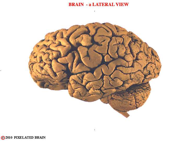
The meninges have been removed and we now look at the lateral surface of the brain. In this view we see the cerebral hemisphere, the brainstem and the cerebellum, but for now we will focus on the hemisphere. Clearly, there has to be a way to refer to different parts of this complex structure. We describe how it is done in the next few figures.
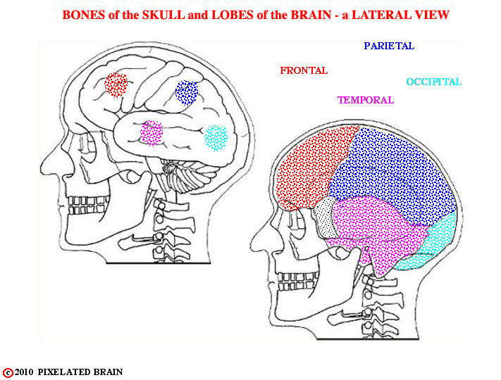
The starting point is to divide the hemisphere into four major lobes, which correspond roughly in name and position to the bones of the overlying skull.
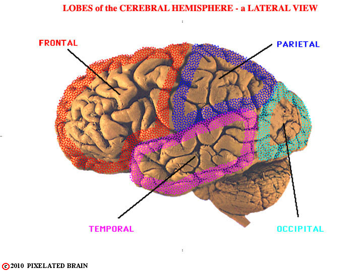
This view shows the general position of the four lobes present on the lateral surface of the hemisphere.
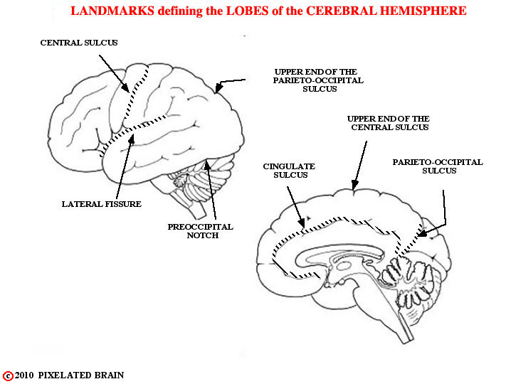
The landmarks used to establish the borders of each lobe are shown here. The central sulcus lies about halfway between frontal and occipital poles and is significantly deeper than adjacent sulci. The lateral fissure differs from a sulcus in that it was created as tissue migrated in an arc-like fashion to form the temporal lobe. The pre occipital notch is a rather predictable indentation in the surface of the brain, and the parieto-occipital sulcus is a very constant and deep sulcus, present on the medial surface of the hemisphere. The anterior border of the occipital lobe is a line drawn between the pre occipital notch and the upper end of the parieto-occipital sulcus.
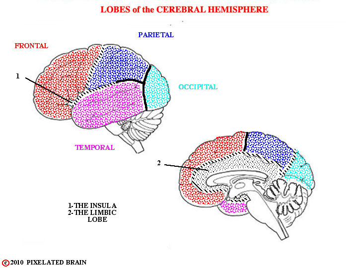
Anteriorly, the lateral fissure separates the temporal lobe from the frontal and parietal lobes; posteriorly, the line separating the temporal and parietal lobes is a somewhat arbitrary extension of the lateral fissure. Because of their unique geometry the insula and the limbic "lobe" to not fit into the four lobe scheme. As the name suggests, the insula is indeed an island of cortex that has been buried by the expanding frontal, parietal and temporal lobes; it shares borders with all three. The limbic "lobe" runs around the medial edge of the hemisphere, sharing borders with all four lobes. Because of its functional importance it is commonly called a lobe, though it hardly seems like one.
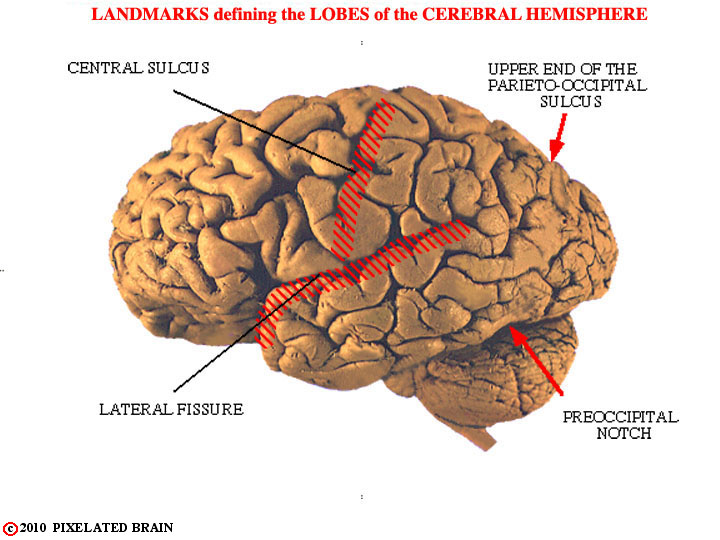
Here are some of these landmarks on a real brain.
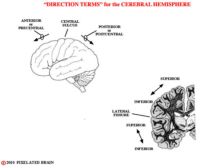
Before describing how each lobe is subdivided into gyri, we need to be clear on the way directions are specified when dealing with the surface of the hemisphere. The most commonly used terms are shown in this view.
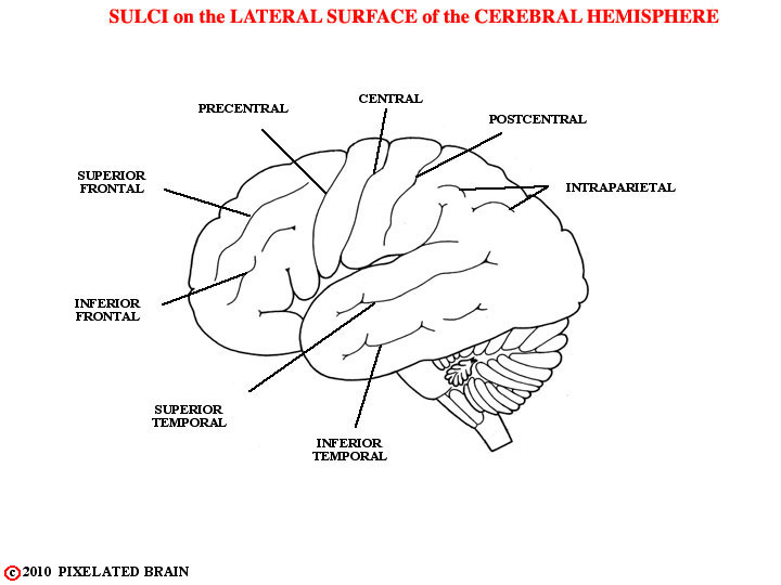
These are the named sulci.
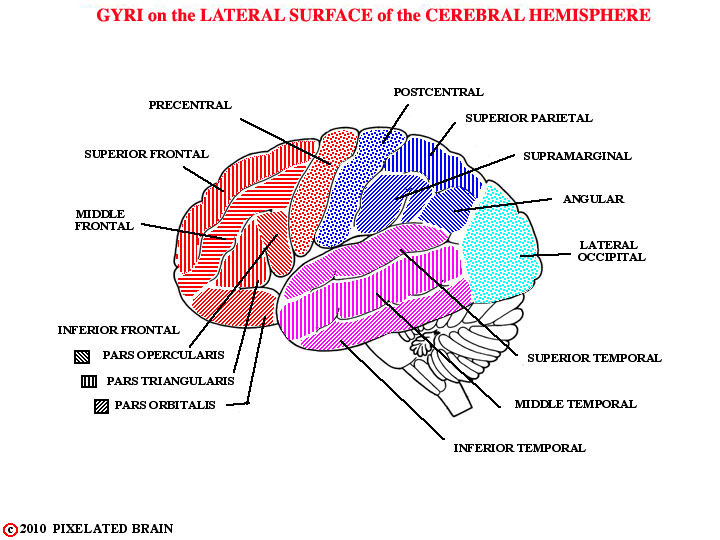
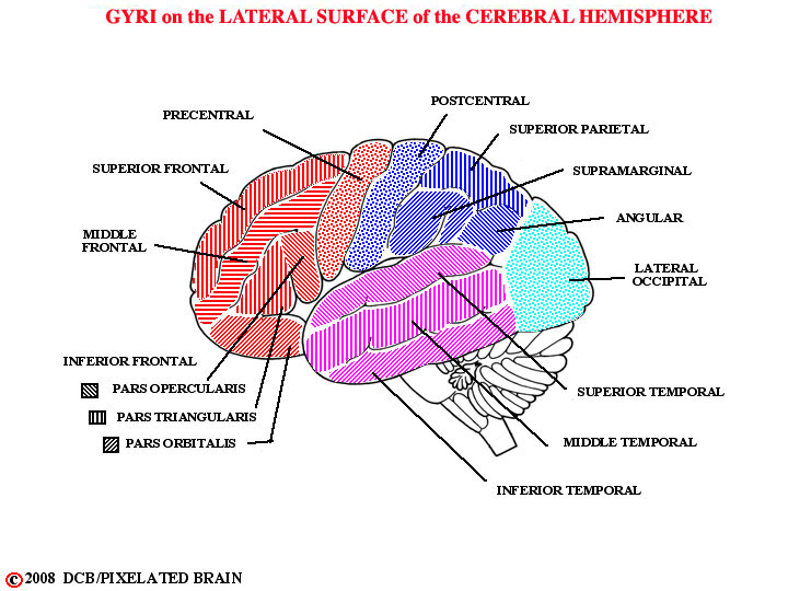
These are the named gyri.
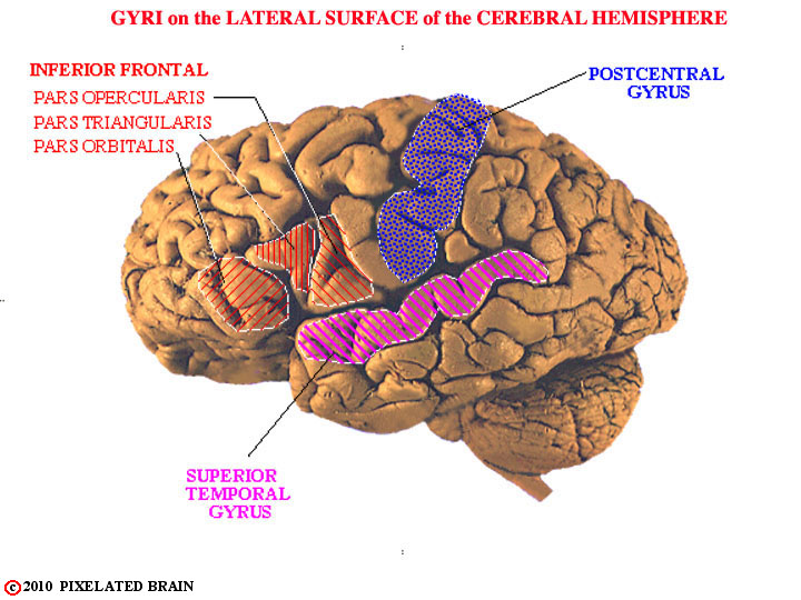
Most textbook figures imply that the pattern of gyri and sulci on the surface of the hemisphere is quite similar from one brain to the next. In reality, there is a great deal of variability between specimens. In the brain we looked at originally, for example, the central, post central and superior temporal sulci are rather clear, with the result that the post central gyrus and the superior temporal gyrus are easily identified, as shown here. In other brains, even the central sulcus (the most obvious of the three) can be difficult to identify.
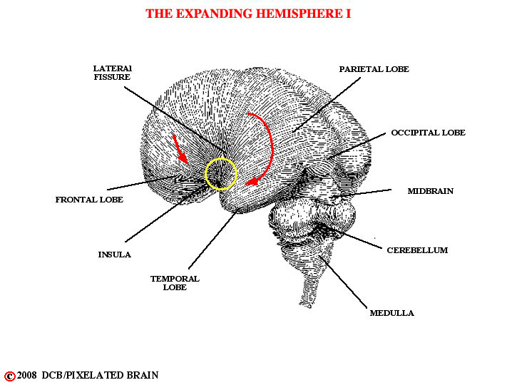
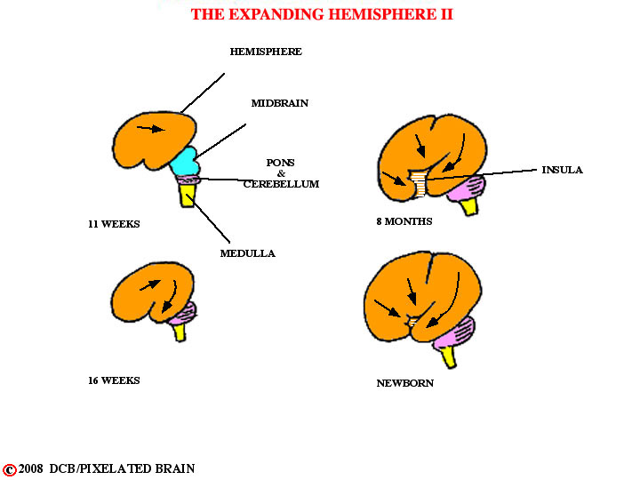
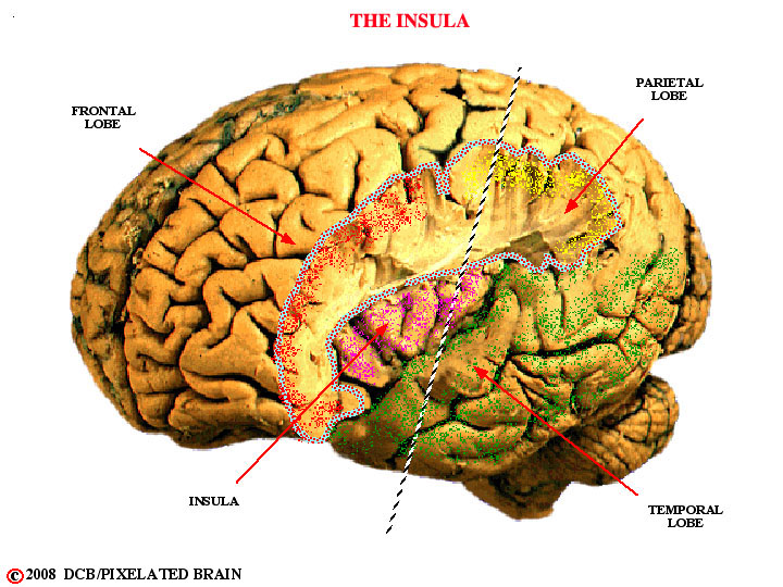
Migration of the frontal, parietal and temporal lobes has tended to bury an "island" of cortex within the depths of the lateral fissure.
Also see Diganat.
Lobe Migration.

If parts of the frontal (red) and parietal (yellow) lobes are cut away this cortical area - the insula (violet) - is revealed, as shown here.
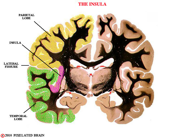
The insula is not truly an island. Its relationship to the lobes of the hemisphere is best revealed in a section such as this one.
For this section, the brain has been cut along the line shown in the previous slide.
For some nice views of the lateral surface of a real brain see the following Digital Anatomist views. Since this is one of the first times in which we have called up these views, we should explain to you how to use them. The Diganat slides are superb views of real brains, but they can be a little overwhelming because the authors have labeled things in great detail. Whenever possible, we want you to call each of them up and then compare to our view. See how many of the structures labeled in our idealized view can be found on the real brain. It's sort of a reality check, and you have to realize that the match won't be perfect.
The left insula.
THE BASAL SURFACE of the BRAIN
The anatomy here becomes a little more complex, because we see not only the basal surface of the hemisphere, but also the surface of the brainstem - the complex region lying between the hemisphere and the spinal cord, and the subject of Module 2. We will focus on the hemisphere, although the brainstem is also shown in this view and the next.
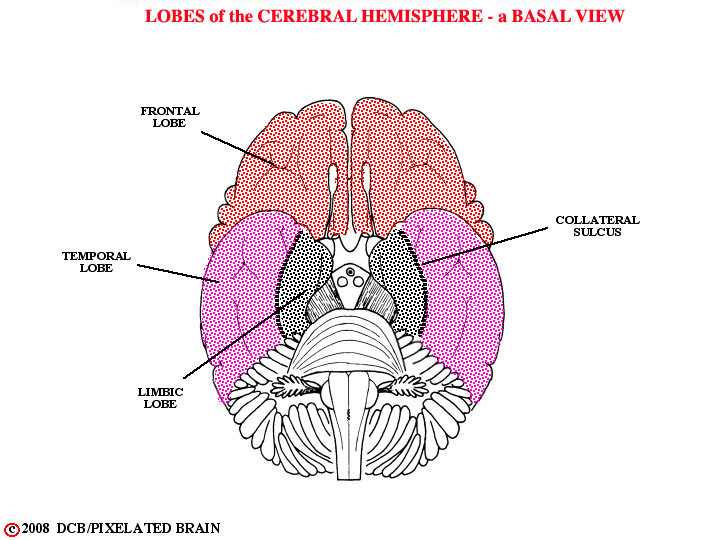
In this view, we identify the lobes of the hemisphere.
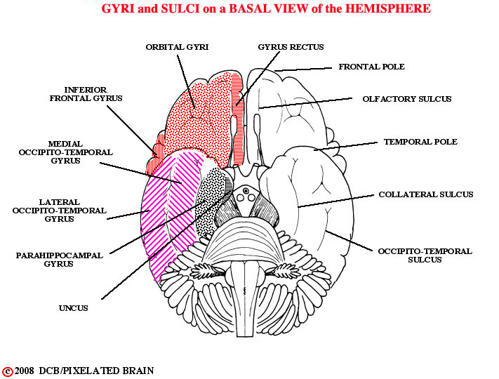
Here, we label the sulci and gyri. The uncus is an important landmark on the medial edge of the temporal lobes.
Compare with.
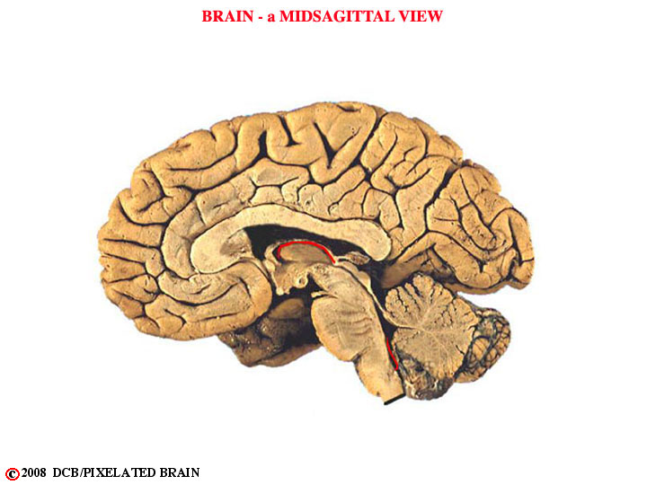
THE MEDIAL SURFACE of the HEMISECTED BRAIN
The medial surface of the hemisected brain is the most interesting of all because it reveals not only another aspect of the hemisphere but also the mid-sagittal surfaces of the diencephalon and brainstem. We will leave the brainstem structures for Module 2 and mainly look at the hemisphere. This is an unlabeled view.
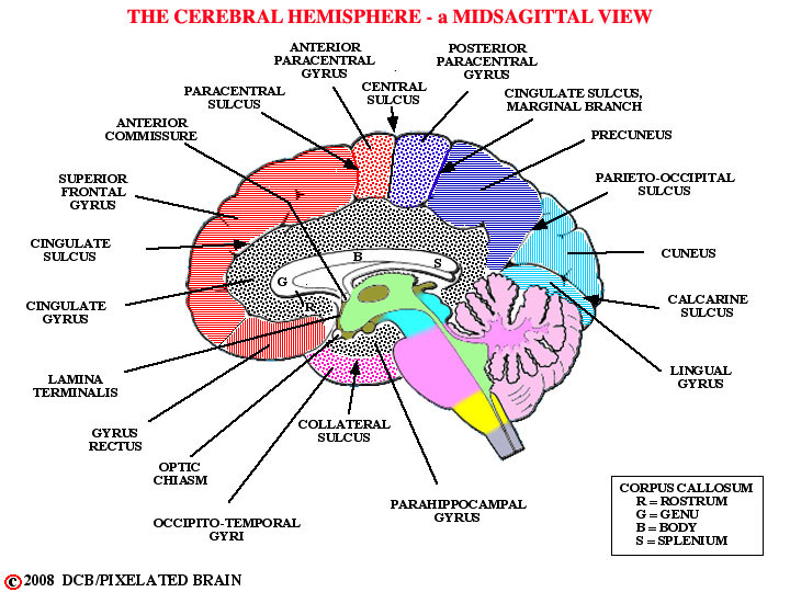
The gyri seen on the medial surface of the hemisphere. Compare with DiganatA_1.4

More anatomy, including some structures we cover in Module 2. Again, compare with DiganatA_1.4
LOCALIZATION of FUNCTION at the CORTICAL LEVEL
At this point, you are asking "Is that it? Do I have all the names I need to describe the surface of the cerebral cortex? If so, now tell me what each of these gyri actually does."
Well, the gyri are a necessary start, but its not that simple (it never is). The gyri are useful landmarks for the neurosurgeon, the neuroradiologist and the neuropathologist and they are used in the reports that these specialists write - operative notes, and the like.
But it turns out that the cellular region lying just deep to the surface of the hemisphere - the light area in the figure- is organized into 6 layers and the thickness and cellular structure of these layers differs from one region to another.
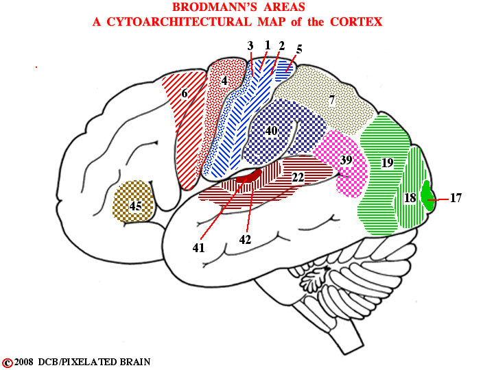
More than 100 years ago, a worker by the name of Brodmann parceled the surface of the cortex out into more than 50 areas, based on the distinctive cellular appearance of each, and his map is used to this day in describing the functional organization of this region.
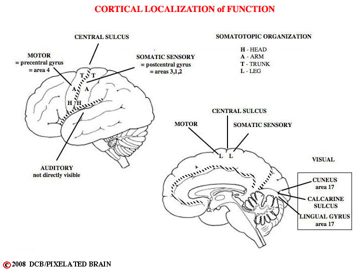
We can now make a few very brief statements about localization of function at the cortical level; scroll back and forth between the 2 figures at the left as you read.
1) The precentral gyrus is the primary motor cortex. Neurons here send descending axons to the spinal cord and brainstem; activity of these neurons results in movement. This area coincides with Brodmann's area 4.
2) The postcentral gyrus is the primary sensory cortex. Neurons here receive a direct input from ascending somatic sensory pathways. This are roughly coincides with Brodmann's areas 3, 1 and 2.
3) The cuneus (actually a gyrus) and the lingual gyrus are the primary visual cortex. Together they receive an input from the retina. This roughly coincide with area 17. 4) The transverse temporal gyri (part of the temporal lobe, but buried in the lateral fissure and hidden from view) are the primary auditory cortex. They receive an input from the ear and coincide with area 41
Many more subtle functions also are carried out in restricted cortical areas.
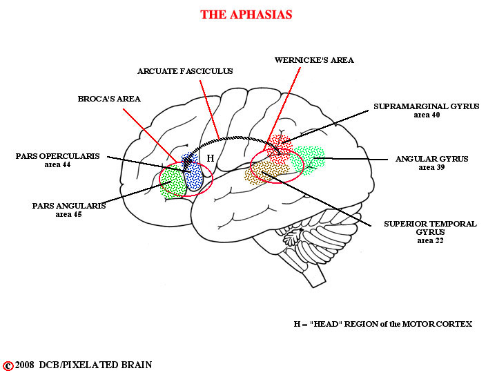
One fascinating example concerns two regions of the brain - Broca's area and Wernicke's area (see figure). Damage of either area in the left hemisphere causes a marked impairment of speech, termed aphasia. Wernicke's aphasia is primarily a difficulty in understanding language. In contrast, subjects with Broca's aphasia understand what they have heard but have difficulty organizing a spoken response. They know what they want to say but are not able to put the words together in a fluent manner. To work properly, Broca's area requires an input from Wernicke's area and the pathway involved - the arcuate fasciculus - is well defined. This history of this discovery is fascinating!
Try to read about it on Pages 9 - 14 in Kandel's 4th Edition.